
UA Little Rock Researcher Receives Grant to Develop More Realistic Model of Human Intestine
Recreating the intricate structure of the human intestine in a laboratory could give scientists a better way to study disease and potential treatments — while reducing their reliance on animal experiments in the process.
With support from a $72,419 grant, University of Arkansas at Little Rock researcher Dr. Sujan Ghosh is working to make that possible by developing a three-dimensional tissue scaffold that more closely mimics the microscopic architecture of the human small intestine.
Ghosh, assistant professor of mechanical engineering at UA Little Rock, received the grant through the Arkansas IDeA Network of Biomedical Research Excellence (INBRE) to develop the bio-inspired model.
The small intestine contains tiny finger-like projections called villi and pockets between them called crypts. These structures play important roles in nutrient absorption, stem cell maintenance, and tissue regeneration, but they are difficult to accurately reproduce for laboratory models.
“A tissue scaffold is essentially a three-dimensional framework that provides cells with a structure to grow on, much like the framework of a building supports its construction,” Ghosh said.
In this project, the scaffold will recreate the villi and crypts to provide intestinal cells with an environment that more closely resembles the one found in the human body. Researchers will then use the scaffolds to support the growth of intestinal organoids, miniature versions of intestinal tissue grown from cells.
Traditional cell cultures are generally grown on flat surfaces, which do not capture the complex three-dimensional structure of the intestine. Animal models can provide greater biological complexity but can be costly, present ethical concerns, and do not always accurately represent human biology.
“Better models will allow researchers to understand how intestinal tissue develops, responds to injury, and regenerates after treatments such as radiation therapy,” Ghosh said. “Ultimately, this work could accelerate discoveries in regenerative medicine and improve how new therapies are developed and tested.”
The grant will support the design, fabrication, and testing of the scaffolds using stereolithography-based 3D printing. As part of the project, Ghosh will optimize the printing process to reproduce the microscopic villus-crypt structures and characterize the scaffolds’ surface morphology, mechanical properties, and structural accuracy.
Researchers will then culture human intestinal organoids on the scaffolds and evaluate how the engineered tissue responds to radiation exposure and how well it is able to regenerate.
Beyond radiation injury, the model could eventually help scientists study conditions such as inflammatory bowel disease and colorectal cancer, as well as evaluate potential drugs and treatments.
More realistic laboratory models could also reduce researchers’ reliance on animal experiments by providing another way to study how human intestinal tissue responds to disease, drugs, and injury.
While the current project focuses on developing and validating the scaffold, the technology could eventually have applications in personalized medicine. Patient-derived cells could potentially be grown on customized scaffolds, allowing researchers to study how an individual’s tissue responds to different treatments.
Dr. Brian Berry, vice provost of research and dean of the Graduate School, said the project reflects both the interdisciplinary nature of research at UA Little Rock and the opportunities that research creates for students.
“This project is a great example of how UA Little Rock researchers are bringing together expertise across disciplines to address complex problems with real-world implications,” Berry said. “Dr. Ghosh’s work not only has the potential to contribute to advances in biomedical research, but it also creates valuable opportunities for students to gain hands-on experience with emerging technologies and research methods.”
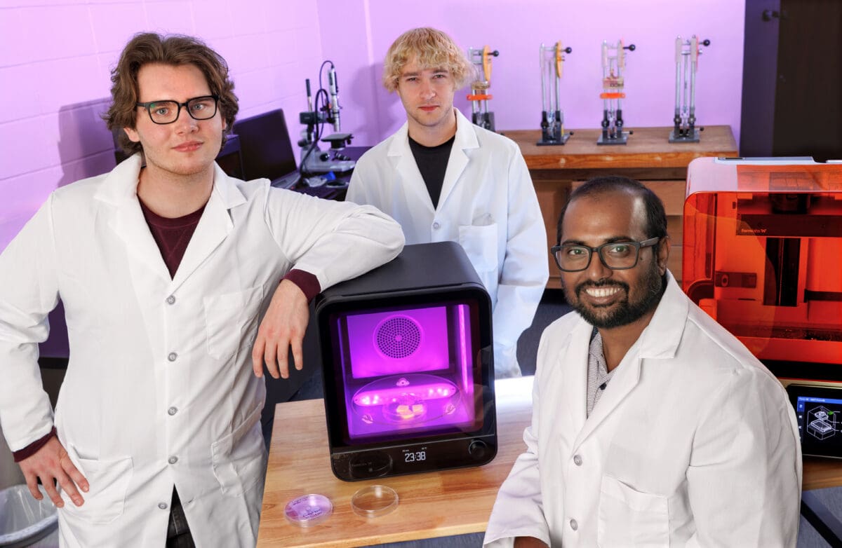
Ben Anderson and William Heap, both mechanical engineering seniors, are already contributing to the project, and they will gain experience in areas such as computer-aided design, high-resolution 3D printing, materials characterization, microscopy, mechanical testing, and biomaterials research.
Anderson is focusing primarily on materials science and additive manufacturing, or 3D printing. While the researchers have successfully reproduced the villus-crypt structures using stereolithography, Anderson is working to create the model through extrusion-based bioprinting. The technique uses organic hydrogel-based materials that can more easily support living cells, an important step toward creating a more biologically realistic tissue model.
For Anderson, the project has also provided an opportunity to explore the intersection of mechanical engineering, biology, and chemistry.
“It is particularly exciting for me to not just use these advanced additive manufacturing techniques, but to learn how to do so competently,” Anderson said. “Performing biomedical engineering work as a mechanical engineer has also been good experience as someone who already has secondary interests in biology and chemistry. Additionally, it allows me to explore interdisciplinary work that I have not done before.”
Heap is approaching the project from a different angle, using computational modeling and computational fluid dynamics (CFD) to study blood flow through the tissue scaffold. His work includes developing simulations and analyzing factors such as velocity and pressure to better understand how the scaffold could perform in a biological environment.
“What excites me the most is being able to apply CFD, an area of mechanical engineering that I have become very interested in, to a research environment like this,” Heap said. “Tissue scaffolds have complex geometries and flow conditions, which makes modeling them a challenging engineering problem.”
Heap said the experience will also help him develop a stronger understanding of computational modeling and fluid dynamics as he prepares for future opportunities in those areas.
Anderson and Heap will also work with biomedical researchers to learn tissue engineering techniques, organoid culture methods, and data analysis, preparing them for careers and graduate study in engineering and biomedical fields.
Ghosh said the opportunity to combine engineering, materials science, and biology is one of the most exciting aspects of the project.
“Advances in additive manufacturing now allow us to recreate biological structures with unprecedented precision, opening new possibilities for developing laboratory models that closely resemble native human tissues,” Ghosh said.
Looking ahead, Ghosh hopes to eventually incorporate additional cell types and biological cues to create increasingly sophisticated intestinal models.